The story of your eyes
Have you ever wondered what’s happening exactly when you have an eye condition? We explain the in’s and out’s of your eye.
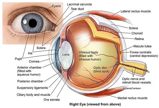
Lens
Transparent tissue that helps bring rays of light to focus on the retina. If the lens becomes cloudy, this is called a cataract.
Presbyopia is caused by the gradual thickening and loss of flexibility of the lens. With less elasticity, the eye has a harder time focusing up close.
Click here for more information on cataracts.
Click here for more information on presbyopia.
Iris & Pupil
The iris is the coloured part of your eye, whereas the pupil is the black part of your eye. They control the amount of light entering your eye by varying the size of the pupil. The larger the pupil size, the more light is let in. This is why your eyes can ‘get used to’ dark environments after a couple of minutes.
Anterior Chamber
Fluid-filled space inside the eye between the iris and the cornea. The amount of fluid exerts a certain eye pressure against the cornea. If the pressure is high or low, this indicates an abnormal amount of fluid in the anterior chamber and can lead to glaucoma (gradual loss of peripheral vision due to damage).
Click here for more information on glaucoma.
Cornea
The transparent front segment of the eye that covers the iris, pupil and anterior chamber. The cornea provides most of the eye’s optical power. When the cornea is scratched, care needs to be taken to ensure that an infection does not infiltrate the eye.
Orthokeratology lenses can gently re-shape the cornea as your sleep to allow clear vision throughout the day without the need for glasses or contact lenses. They have the added benefit of controlling progressive myopia.
Click here for more information on orthokeratology lenses.
Ciliary Body & Muscles
Responsible for focusing the lens to help you see clearly at different distances
Retina
The retina is the “camera” of your eyes that tells the brain what you are seeing. Its structural integrity is important in making sure light can focus onto it properly in order to do this.
Click here for more information on retinal tears and detachment.
Click here for more information on floaters and flashes.
Macula Lutea
The area of the retina that is responsible for sharp, detailed vision, and allows you to recognize faces, drive, read, and many other daily tasks. Although the macula is a very small area in the retina, it is actually responsible for 80% of your vision! Macular degeneration occurs when deposits accumulate underneath the retina and leads to a painless loss of vision.
Click here for more information on macular degeneration.
Sclera
The white protective outer layer of the eye. When you experience eye irritation or infection, this area can appear red and acts as a signal for help!
Click here for more information on dry eye.
Choroid
The layer of the eye that lies between the retina and sclera. This layer provides nourishment to the outer layers of the retina.
Vitreous
Transparent, jelly-like material that fills the interior of the eyeball. As you age, the vitreous can start to degenerate and form floaters in your eye. Floaters aren’t harmful, but can be annoying.
If you’re concerned with any possible eye conditions, please don’t hesitate to contact us on 3345 3383 for a comprehensive eye test!
The Plumbing System
Problems in this area is the main cause for dry eyes.
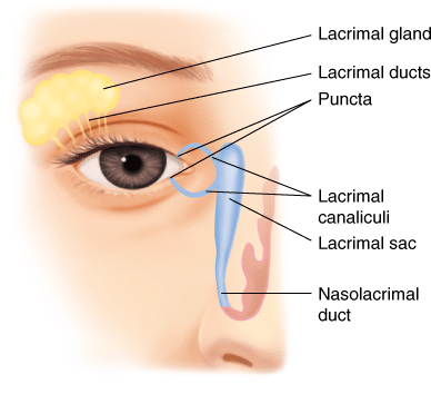
Lacrimal Gland
These are responsible for the production of healthy tears to protect the surface of the eye. Aqueous deficient dry eye is a form of dry eye where the tear production is reduced. This is more commonly associated with Sjogren’s syndrome.
Click here for more information on dry eye.
Puncta
These act as the drainage system for the eye. In cases of aqueous deficient dry eye, we can also offer a treatment called punctal plug to block the duct and reduce the drainage.
Click here for more information on dry eye.
Lacrimal canaliculi/Lacrimal sac
In some cases, the lacrimal passage can be blocked and result in watery eyes from reduced tear drainage. An in-office treatment called a lacrimal lavage can be done to flush out any debris and blockages.
Click here for more information on dry eye.
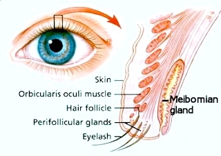
Meibomian gland
These produce healthy oils that prevent your tears from evaporating too quickly. Meibomian gland dysfunction (MGD) is the most common cause for dry eye, but luckily it is easy to maintain if you know how!
Click here for more information on dry eye.
Ask us about our comprehensive dry eye assessment to find out what’s causing your dry eyes, and what you can do about it. Click here for more information on dry eyes.