Retinal Tears or Detachment
If you have noticed sudden flashes, floaters, or vision loss,
call us on 07 3345 3383 or book online today!
The retina is the “camera” of your eyes that tells the brain what you are seeing. Its structural integrity is important in making sure light can focus onto it properly in order to do this. However, sometimes damage to this layer at the back of your eye can results in tears or complete detachment of the layer, resulting in sudden vision loss and increased presence of flashes and floaters. If not treated quickly, retinal tears and detachments can lead to permanent vision loss.
The retina
The retina is a thin layer of tissue that lines the back of the eye, the closest layer to the vitreous gel-like fluid that fills the eyeball. It is made up of millions of light-sensitive receptor cells and nerve fibres that convert light from the environment you are seeing into neural signals that are sent to the brain for visual processing.

Image: An opthalmogram, an image of the fundus of a healthy human eye.
The signs of retinal tears or detachment can range from subtle flashes and floaters to sudden blindness, and if only a small area is affected, you may not experience any symptoms. This is why it is very important to not ignore abnormal visual changes and seek advice from an eye care professional, especially if you have been exposed to any risk factors like listed on this page.

Signs of retinal tears of detachment
- Sudden increase in floaters. Floaters naturally increase with age and are not always a sign of retinal tears or detachment. However, sudden onset and increased in size and number of floaters should be checked
- Sudden flashes of light
- Shadow / grey curtain in your peripheral (side) vision that can move towards your central vision
- Sudden decreased or blurry vision
It is very important to not ignore symptoms of retinal tears and detachments and seek advice from an eye care professional. If you have experienced any of these symptoms, please call (07) 3345 3383 to book an assessment.
Risk factors
- Age. The vitreous fluid shrinks with age, sometimes pulling the retina with it.
- High myopia / short-sightedness. Myopic eyeballs tend to be longer and can stretch the retina.
- Thin areas of retina. Some eyes have thinner patches of retina which increase the risk of tears and detachments. These are often picked up during eye examinations.
- Eye injury. Trauma to the eye can cause damage to the retina. Especially when safety eye wear is not worn in high-contact sports, construction work, etc.
- Eye surgery. Invasive eye surgeries may put the retina at risk.
- Previous retinal tears or detachment.
- Family history of retinal tears or detachment.
What happens if I don’t treat retinal tears or detachment?
If the retinal tear or detachment isn’t treated, more of the retina can detach which increases the risk of permanent vision loss. Retinal detachment of the entire retina causes permanent blindness.
Early detection
Retinal tears and detachments require urgent assessment. The earlier retinal tears and detachments are detected, the lower the risk of permanent vision loss.
Our optometrists are equipped for early detection of retinal tears and detachments with the following techniques and instruments:
- Slit lamp microscope to look inside the back of the eye and view the retina
- Dilating eye drops to enlarge the pupil and view much more of the peripheral retina which sometimes can’t be seen with normal viewing techniques
- Digital retinal photographs provide a detailed image of the central retina
- OCT imaging is a non-invasive ultrasound scan that assesses the retinal layers and shows any retinal lifting or damage.
If detected, the optometrist will quickly send you to an ophthalmologist.
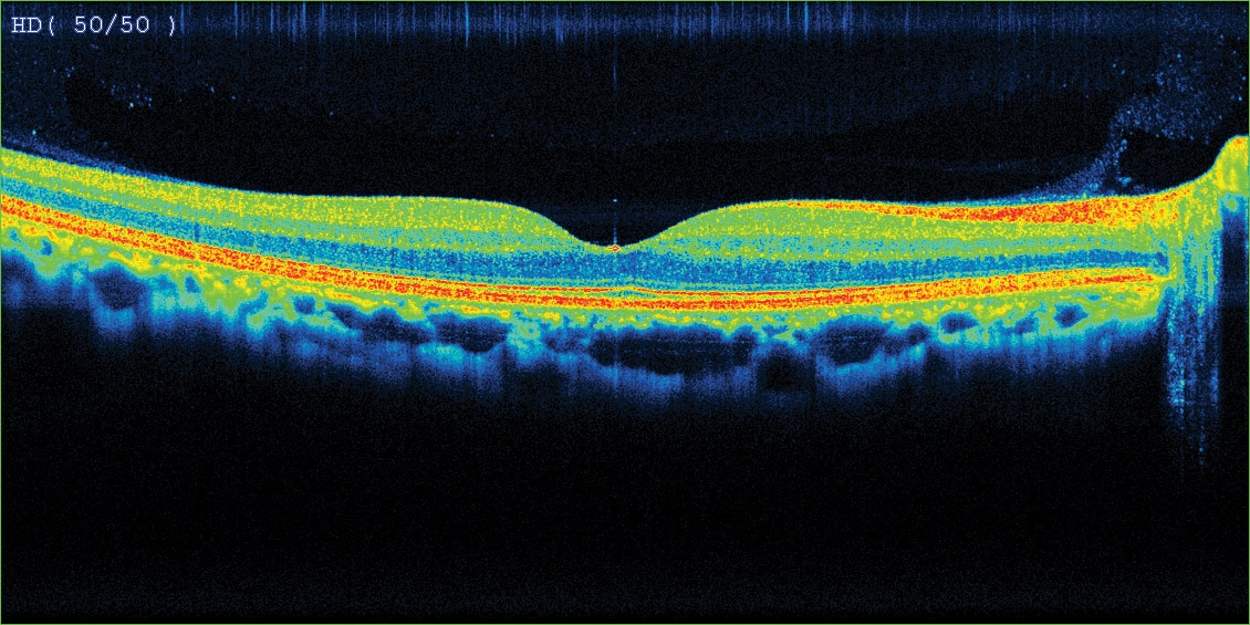
Left: OCT scan of normal macula
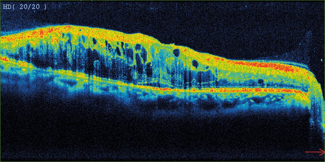
Right: OCT scan of diabetic macular oedema
Treatment options
- Laser treatment. Seals down the retina around the tear or detachment so it doesn’t spread
- Vitrectomy. Replaces the vitreous fluid with saline to prevent further pulling on the retina for large tears.
- Scleral buckle. Silicone band is wrapped around the white of the eye to push the eye towards the tear or detachment until it heals.
- Pneumatic retinopexy. A tiny gas bubble is injected in vitreous fluid to put pressure against the tear or detachment until it heals.
Approximately 85% of retinal detachments can be surgically reattached if diagnosed early. The amount of vision restored after surgery will depend, however about 40% of people with successful surgery will have excellent vision.