Our Technology
Quicklinks
Ultra-Widefield Retinal Imaging
Personalised lens measurement device
Ultrasound to allow early detection of a range of eye diseases.
Ultrasound to monitor progressing myopia, cataracts, glaucoma
Diagnosis and monitoring glaucoma and other optic nerve diseases
Advanced dry eye assessment
Eye curvature for custom contact lenses
Computerised eye movement tracker when reading
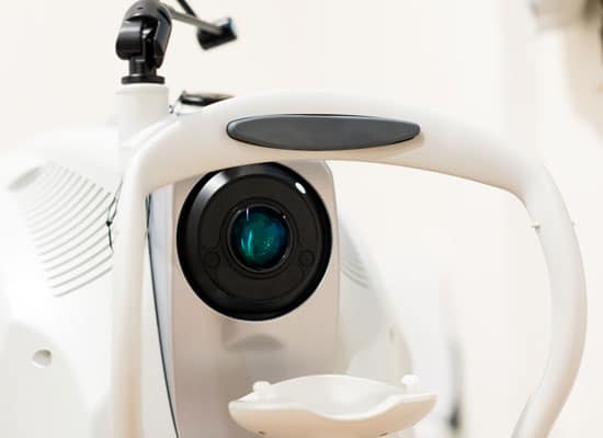
Optomap
Optomap is the latest technology in eye care. A high-tech imaging tool that allows us to see and image virtually all of your retina in a single shot.
A comprehensive view that was previously unavailable, allows us to better diagnose & assess your ocular and overall systemic health.
Optomap in a single, quick shot, produces photos or angiograms of about 82% of the retina. These optomap images provide superior visibility of the retinal periphery allowing us to see, document, show you, and follow pathology that could not be seen with traditional eye cameras.

Book an appointment for a comprehensive evaluation of your retinal health using the latest Optomap technology.

Optomap will show us if you have any signs or conditions such as Diabetic Retinopathy, Hypertensive Retinopathy, Retinal Holes, Tears and Detachments, Macular Degeneration, Malignant Melanomas or other retinal disorders.
Most eye disorders do not cause pain or other symptoms and when detected early enough, many retinal problems can be treated to avoid vision loss. However, once a retinal problem has caused a loss in vision, it is almost never reversible. Thus, early detection is vital to preserving good eye health.
Snapshot summary
● What: Detects any retinal disorders.
● When: Eye examination or if you have any concerns.
● How: Simply look into the device one eye at a time and the image of your retina will be taken. It’s fast, painless, and comfortable. Nothing touches your eye at any time*.
● What’s new: Produces photos or angiograms of about 82% of the retina, compared to 20% previously using traditional ophthalmic cameras.
● Duration: < 0.5 second per eye for an optomap image.
*Under normal circumstances, dilation drops might not be necessary, but your eye care practitioner will decide if your pupils need to be dilated depending on the health of your eyes.

Visioffice 3
Visioffice 3 technology offerd personalised products to the highest extent, making the lenses adapt to you and not the other way around.
Snapshot summary
- What: Personalised spectacle lens measurement device
- When: Personalised spectacle lens measurement device
- How: In-store capture with a HD capture and Activisu®’s 3D reconstruction technology
- Duration: 5-10 minutes
Click here for more information on how the Visioffice 3 personalises your lenses.
Premium Optical Coherence Tomography (OCT Imaging)
OCT Imaging is a technique used for obtaining a detailed image of the retina and the multiple layers underneath. This is not possible with the standard eye examination which only allows us to view the surface of the retina. This is important because many eye diseases often begin within or underneath the retina.
It is effectively an ‘optical ultrasound’, using imagine reflections from within the eye using light waves to provide cross-sectional images. OCT provides more information than other imaging methods such as ultrasound or MRI.
Benefits include:
- Early detection of a variety of eye diseases. This results in a significantly better chance of retaining your vision as compared to detection at a normal eye exam.
- Fast
- Non-invasive with no eye contact*
- No radiation
Premium Features:
- Ultra high resolution
Active gaze tracking allows the ability to perform 100 scans in the exact same position producing high resolution low noise scans. This is superior to standard machines which do not take into account head movements and may produce lower quality scans.
- Accurate results for progression diagnosis
‘Eye tracer’ locks onto the retina and guarantees that scans done on subsequent visits are performed in exactly the same position, time after time. This maximizes eye disease detection and accurate progression analysis. This enhances early detection and treatment
Should I have an OCT scan and how often?
We recommend everyone, including those with healthy eyes, to have an OCT scan performed. This will establish a baseline, so when your eyes start to change in the future, we can compare what has changed and by how much.
After performing a baseline OCT, we recommend OCT imaging every three years with your standard eye test. However if you are over 40 years old or are experiencing any eyesight problems, a yearly scan is preferable. The images are stored electronically, so regular OCT imaging helps us to build a clear picture of your eye health over time. By comparing records, it’s easier to identify subtle changes that may indicate the early signs of disease.

Snapshot summary
- What: Provides earlier detection of nerve damage compared to visual field tests. Can pick up glaucoma up to 8 years before noticing symptoms.
- When: Separate follow-up exam. Included in our ‘Comprehensive glaucoma assessment package’.
- How: Non-invasive ultrasound scan to assess the volume of the nerve fibres supplying the optic nerve head
- Duration: 5 mins per eye
Specific indications to perform an OCT scan include:
- Patients with unexplained vision deterioration
- Abnormality detected at the back of the eye during a routine eye examination
- Abnormality detected during a retinal photography scan
- Patients with diabetes (who have an increased risk of developing retinal disease)
- Patients with a family history or other increased risk of developing macular disease** or glaucoma^
- Patients with high intra-ocular pressure
- Patients with visual field loss
- Evaluation of corneal conditions and anterior chamber depth
** The macula is a specialised area of the retina responsible for the detailed vision necessary for everyday tasks such as reading and recognising a person’s face. An OCT scan is usually the best method to detect and monitor a range of macular conditions including macular degeneration, macula traction, epiretinal membrane, macular hole, diabetic retinopathy and oedema.
^ Glaucoma is a disease resulting in damage to the optic nerve (the nerve bundle which connects the eye to the brain). Increased pressure in the eye can lead to damage to the optic nerve. As damaged nerve fibres cannot regenerate, early detection and treatment is vital to prevent further nerve damage. The OCT measures the thickness of the nerve layer around the optic nerve. Efficiency of the drainage system in the front chamber of the eye and thickness of the cornea are also contributing factors to glaucoma. The OCT provides information on these factors.

Snapshot summary
- What: Detects sensitivity of your field of vision.
- When: Separate follow-up exam. Included in our ‘Comprehensive glaucoma assessment package’.
- How: Small spots of light are presented to test and measure sensitivity of vision at different areas around your visual field.
- What’s new: Faster = More comfortable testing experience.
Increased sensitivity = Diseases are detected even earlier and easier. - Duration: 3-4 mins per eye (50% faster than previous testing methods)
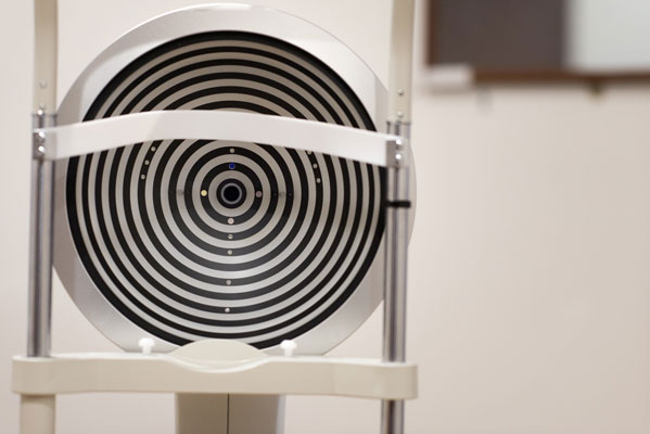
Visual Field Test
The visual field test is a subjective measure of central and peripheral vision, or “side vision,” The use of visual field test is essential in diagnosing and monitoring glaucoma, or any vision loss as a result of damage to the visual pathways of the brain or other optic nerve diseases. It uses a light flashes that is repeatedly presented in different areas of your peripheral vision. A visual field test is performed as soon as glaucoma is suspected, and the visual field data is used to determine the severity of disease. This staging information is useful in determining follow-up and/or treatment strategies.
OCT and retinal photograph imaging of the optic nerve and surrounding tissues is an objective test that can also detect glaucoma damage and progression. However, at this time it has not replaced the visual field test. We still need both types of testing because there are times that the optic nerve changes before the visual field, but also times when changes to the visual field are observed before damage to the optic nerve is detected.
Snapshot summary
- What: Detailed assessment of dry eyes
- When: Included in our ‘Dry eye assessment package’.
- How: Various scans
- Duration: 15 mins
Oculus Keratograph 5 (K5)
This machine has revolutionised Dry Eye assessment. The K5 allows us to have an in-depth look at the eyelid anatomy and tear film to diagnose and manage dry eyes quickly and accurately. It allows us to scan and measure critical features such as:
- The quantity of tears,
- The quality of lipids in tears,
- Tear break-up time,
- The integrity of the Meibomian Glands.
The K5 has the following features:
- Meibo-scan allows us to see the structural changes of the glands
- TF-scan uses infra-red illumination to assess the quality and quantity of the tear film.
- Infra-red illumination evaluates the tear film.
- Lipid layer thickness which directly links to tear evaporation and dry eye symptoms. If the lipid layer is too thin or absent, the tear evaporation rate and the tear film instability increases.
- R-scan is the first and only technology that objectively measures and classifies the degree of redness in blood vessels.
Corneal Topographer
A corneal topographer is a non-invasive instrument which maps the curvature of the cornea and outer-part of the eye. Corneal topography is essential in allowing optometrists to fit custom contact lenses and diagnose eye conditions such as keratoconus, Terrien’s marginal degeneration, or monitor pterygium.

Snapshot summary
- What: Provides data to measure and predict myopia progression
- When: Every 4-6 months for patients undergoing myopia management treatment
- How: Ultrasound scan
- Duration: 2mins per eye
AL Scan
Assists those with cataracts and progressing myopia.
Because protecting children’s vision is so important to us, we have invested in training and equipment to provide the best solutions for myopia. This equipment is rare among Brisbane optometrists and can measure exact eyeball length to the closest 0.05mm. It will provide valuable information to measure and predict myopia progression, and to determine the most suitable course of action.
It allows:
- Pre-emptive detection of myopia progression in young eyes – allowing for optimum intervention strategies
- Measurement of cataract density for more detailed referrals to specialists
- Assessment of the Intraocular Cataract Implants (IOL) post-surgery
- Provides valuable information for higher accuracy glaucoma detection
- Improved accuracy of post-operative glasses prescriptions
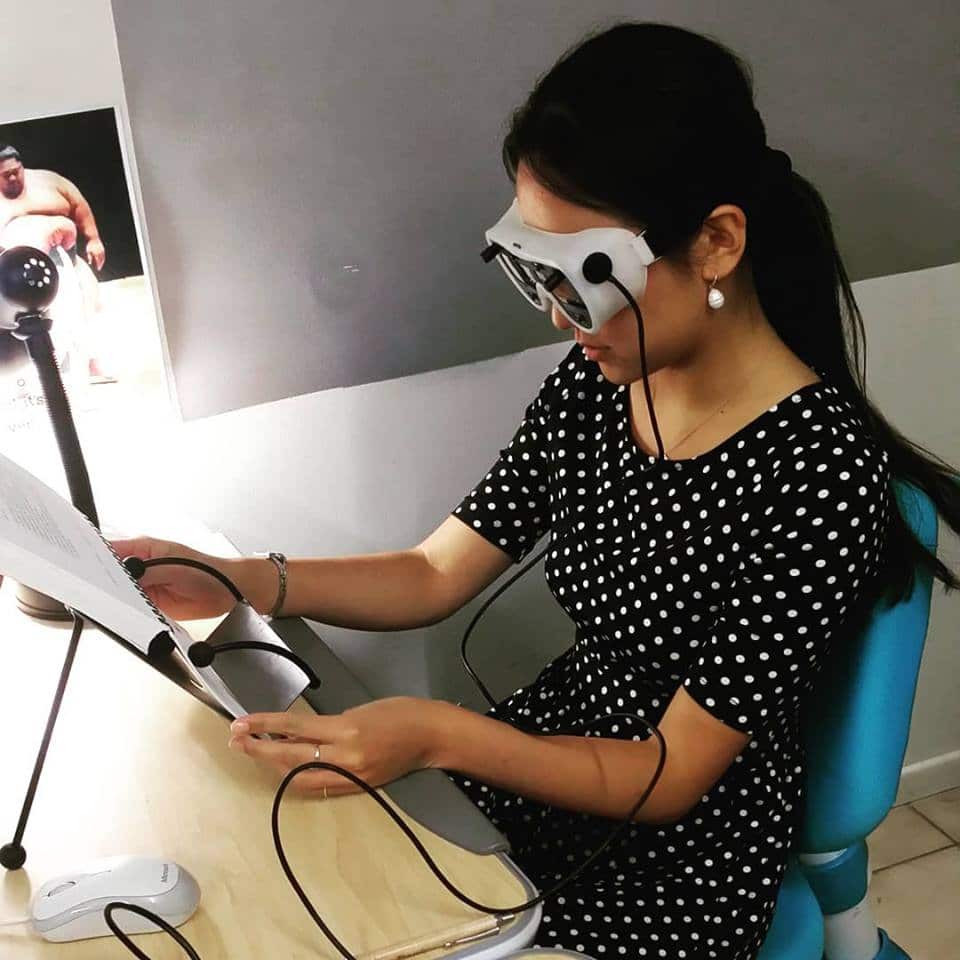
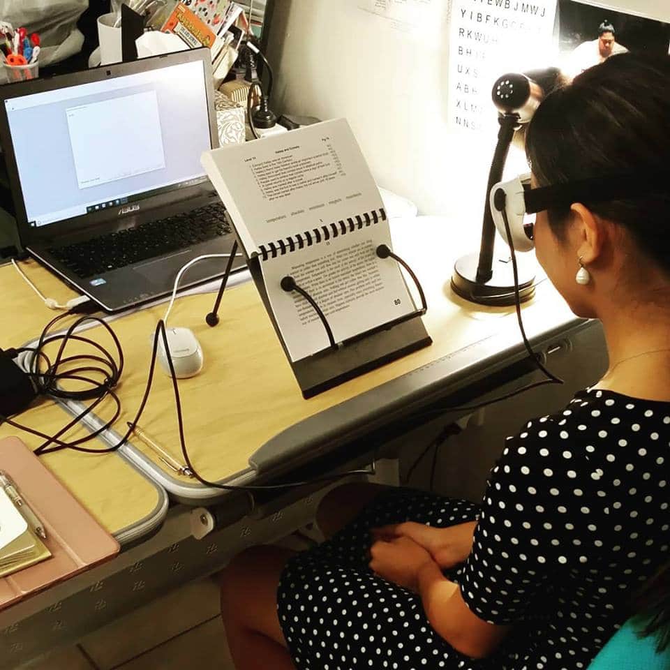
Readalyzer – Computerised eye movement tracker
Do you or your child have trouble with reading no matter how hard they try?
Problems with eye movements can result in:
- Loss of place when reading
- Skipping and omitting words
- Skipping lines or sentences
- Difficulty with copying
- Sore eyes
- Poor hand writing and spacing between letters
- Poor hand eye coordination
- Changed body posture so that eyes are excessively close to the page when reading or writing.
Eye movements require the highest level of precision in the human body. But just like any other muscle, eyes can be trained to improve performance and reading ability.
Call 3345 3383 to book an appointment for a Readalyzer assessment.
This video is a reading simulation of our very own optometrist, Lucy – not a result you would expect from an accomplished professional! Although the struggle is almost palpable, it just goes to show how hard work and perseverence can overcome the highest hurdles.
Grace & Vision Optometry accepts all major health funds
Use your optical health funds benefits on a new pair of prescription glasses or sunglasses.

... and many more. Feel free to contact us if you don't see your health fund.